0 Cart
Added to Cart
    You have items in your cart
    You have 1 item in your cart
    Total

    News

    Researchers find a "consciousness switch" deep in the brain

    There's an incredible amount we don't understand about the workings of the human body and brain, and consciousness itself remains one of the great mysteries of science. Which is weird, because in some senses it's about the only thing we can be sure exists. It doesn't matter whether life is a simulation, or even whether you really exist, I know that I'm having a subjective experience, and that may be the only truth each of us can be sure of.

    The fact that this subjective experience can be switched off, whether by dropping into a deep sleep, getting knocked out or going under anesthesia, does nothing but add to the weirdness of it all. Things are happening to and around you, your awareness is just not online. The fact that there are highly paid and highly trained anesthetists who put people to sleep for a living merely reflects decades of trial and error, rather than a complete understanding of how an anesthetic drug works.

    Now, researchers from the University of Wisconsin, Madison, appear to have made a bit of a breakthrough. In a study published in the journal Neuron, the team showed that at a frequency of 50 Hz, electrical stimulation of the central lateral thalamus, a region once thought of mainly as a relay, amplification and processing station, was able to pull macaque monkeys out of an anesthetized state and elicit normal waking behaviors.

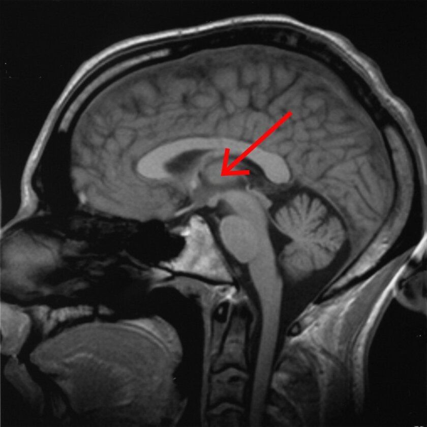
    Scientists have long been studying the thalamus, which sits deep in the brain near the brain stem, to learn what role it plays in sleep, waking, consciousness and alertness. But this research, in which targeted electrical stimulation was applied to a specific area, narrowed the search down further than ever before. The electrodes used in the study were more tailored to the shape of the brain structures they were designed to work on, and the electrical stimulation was designed to mimic the activity of a normal, waking brain.

    The central lateral thalamus is found deep in the center of the brain, close to the brain stem

    The central lateral thalamus is found deep in the center of the brain, close to the brain stem AxelBoldt/Wikimedia Commons

    "We found that when we stimulated this tiny little brain area, we could wake the animals up and reinstate all the neural activity that you'd normally see in the cortex during wakefulness," says senior author and assistant professor Yuri Saalmann. "They acted just as they would if they were awake. When we switched off the stimulation, the animals went straight back to being unconscious."

    The researchers hope this discovery might be able to help people with "disorders of consciousness" – for example, it might be possible to bring people out of comas with consciousness-starting devices, or give narcolepsy sufferers the ability to self-stimulate when they're falling asleep at an inopportune time.

    What's more, anesthetists could use these findings to potentially keep tabs on whether patients are properly under, and when they might be starting to wake up, avoiding some rare but traumatic operating theater experiences. You've also got to wonder if there's a cure for insomnia in there somewhere, or the ability to switch off and sleep through a long plane flight. Time will tell.

    The study was published in the journal Neuron.

    Source: Cell Press via Science Daily

    By Loz Blain


    A Guide to How the Brain Works

    If the human body was compared to a machine, the brain would be the computerized system that receives input and controls what all the other parts do. In comparison to other animals, the human brain is small, yet often much more complex. Even with larger brain sizes, most animals do not have the complexity levels that the human brain does. Although the brain can be divided into multiple sections, they all function as one whole. All areas rely on the other areas to perform both complicated and simple tasks, as well as voluntary and involuntary tasks.


    Basic Characteristics

    The average brain weighs between 2.8 and three pounds and is about 15 centimeters long. It has hundreds of wrinkles and folds on the surface, compacted into an oval shape. The brain is covered by layers of membrane called the meninges and is encased by the skull.


    Although this seemingly mushy mass of folds might appear simplistic, it is quite complex. With this oval mound of tissue, there are billions of neurons, dendrites, and axons with trillions of connecting synapses. The brain also has glands and other smaller structures, divided into 3 main sections- the cerebellum, cerebrum, and brain stem.


    Cerebrum

    The cerebrum is the top section of the brain, and it is divided into two hemispheres, sometimes called the left and right brains. Both the hemispheres manage the opposite side of the body, and four lobes are contained within these two hemispheres, duplicated for each hemisphere. The cerebrum has two layers called white and gray matter, denoting the inner and outer layers, respectively. The gray matter is the cerebral cortex, while the white matter is called the cerebral medulla.


    It is in this section of the brain where planning and organizing, intelligence, and motor functions primarily occur. The processing and understanding of language occur here as well. Each of the four lobes has its own purpose yet work together to handle the tasks controlled by this area of the brain.


    Frontal Lobes

    In comparison to the other three, the frontal lobe is much larger. Located at the front of the brain, this lobe is responsible for several important things. Essentially, it is where information is processed into physical movement. Although the frontal lobes of both hemispheres overlap in their functions, the right and left frontal lobes do have a few different functions.


    The right frontal lobe tends to have more involvement in distinguishing specific aspects of communication, including distinguishing between negative and positive facial expressions. This lobe is also believed to distinguish certain auditory cues in speech, such as tone and emotional values in a person’s voice. Studies have also shown that the right side of the frontal lobe has more electrical activity during the expression of negative emotions. In contrast, the left frontal lobe predominantly handles language related movements, such as body language and physical emotional expression.


    One thing the frontal lobe controls is motor function, and this lobe also controls speech, reasoning, and problem-solving. Emotions and memory are managed by the frontal lobe, and in addition, eye and skeletal movements, judgment, and personality are handled here as well.


    Movement tasks are processed by a portion of the frontal lobe called the motor cortex. The motor cortex is divided into three areas- the primary motor cortex (M1), the premotor cortex, and the supplementary motor area. Controlling most of the movement, the primary motor cortex is responsible for the force, speed, and direction of voluntary movements. The premotor cortex determines which muscles should be used for movement, while the supplementary motor area manages the coordination of simultaneous smaller movements required to perform larger movements.


    Parietal Lobes

    Located near the middle of the brain, the parietal lobe has the primary function of managing sensory information. Pain, taste, and touch sensations are processed in this lobe, and in addition, spatial orientation, writing, and some aspects of speech are also managed by the parietal lobe. Although most sensory information processing is spread between other lobes, the parietal lobe is the main controlling lobe.


    In addition to sensory processing, the parietal lobe is responsible for visuospatial navigating and proprioception through the posterior parietal cortex. It can also help distinguish the number of objects in a visual field and coordination of attention, managed primarily by the superior parietal lobule. Known as Geschwind's territory, the inferior parietal lobule aids in processing language and facial expressions.


    Temporal Lobes

    Located below the other lobes, the temporal lobes manage processing for sensory and auditory functions, speech related tasks, and memory storage. The temporal lobes also control facial recognition and emotional responses. Within the temporal lobes, there are a few key areas- Wernicke’s Area and the amygdala- that help with functions. The Wernicke’s Area assists in processing words and interpreting speech, and the amygdala assists in processing memories and emotions by receiving sensory information from areas in the cerebral cortex including the thalamus.


    Occipital Lobes

    Located near the back section of the brain, the occipital lobe primary handles vision related tasks. All information relayed by the eyes is processed in this lobe, including the ability to read. The occipital lobe also works to send information about images such as the shape, size, and color and it also aids in spatial recognition tasks that include assessing the depth and distance of objects.


    Cerebellum

    The lower portion of the brain is called the cerebellum, and its main duties include controlling balance and coordination. The cerebellum has a role in movement, but only in the sense of making movements more fluid. Generally, the cerebellum helps more with fine motor skills, which includes actions that require practice. It comprises about 10 percent of the brain’s weight but contains more than half of its neurons.


    As with the cerebral cortex, the cerebellum also has folds that contain gray and white matter, and the folds are much more compact and smaller than others. It has three lobes that have the primary responsibility of receiving information from specific areas of the body. The anterior lobe is responsible for receiving and processing information sent by the spinal cord. Information from the cerebral cortex and brainstem are received by the posterior lobe. The flocculonodular lobe processes information sent by the vestibular nerve.


    Brain Stem

    Important functions, such as heartbeat and breathing, are controlled by structures in the brain stem. Sitting just below the limbic system, the brain stem connects to the spinal cord. Ultimately, most information processed by the brain is sent to the neural pathways of the spinal cord. The pons, midbrain, and medulla oblongata are structures found in the brain stem.


    Functioning with dual duty, the midbrain processes visual and auditory information, while also controlling movement. The pons is the largest section of the brain stem and joins part of the cerebral cortex to the cerebellum and medulla oblongata. It plays a large role in breathing and sleeping. Acting as the main control center for the lungs and heart, the medulla oblongata regulates blood pressure, breathing, swallowing, and heart rate.


    Working Together

    All aspects of the brain work together through the nervous system. Each region relies on the others, working to maintain homeostasis. Neurons, dendrites, and axons run throughout the entire brain in a system of neural pathways. Information is sent through these pathways in the form of electrical impulses. There are two parts of the nervous system, each responsible for different things.


    Information is received from the body and sent through a network of pathways to the appropriate processing center or lobe. The information is then sent across other areas if needed, to process the appropriate response. At this point, the response information is then sent back through the pathways to the muscles and organs required for an activity. For example, if something hot is touched, this information is sent from the nerves in the hand to the brain. The sensation is processed by the lobes that handle sensory information and then the response to jerk away from the heat is sent back to the hand to avoid more pain. All this back and forth transmission is done in a matter of seconds.


    As the main control center, the central nervous system (CNS) contains the spinal cord and brain, while the peripheral nervous system (PNS) connects the CNS to the rest of the body via nerve pathways. All sensory information is received and processed in the brain and then dispersed to the appropriate neural pathways around the body. Each system is completely dependent on the other for the body to function properly.


    As technology advances, medical professionals can learn more about the brain and the process behind how it works. Through imaging tests, for example, doctors can view electrical activities occurring within the brain and distinguish between damaged and healthy tissue. Understanding the brain and its complex functions allow an insight into many problems, including several psychological and processing disorders, and it can also provide an insight into the basic functioning and learning process. The human brain might be small in comparison, but it is one of the most complex organs in the body.

    Everything You Need to Know about Functional Supplements

    Functional supplements are taken by people to fill a specific nutrient deficiency or improve a distinct area of their health. Types of functional supplements vary and may include detox supplements, digestion supplements, focus supplements, immunology supplements, and sleep supplements. For functional supplements, many benefits may exist including a lower risk of medical conditions, improvements to health, and an increase in brain function.*  When taking functional supplements, you should follow the directions on the supplement package and research any ingredients listed on the label that you are unsure of. Take time to learn about functional supplements and how they can benefit your health.

    Brain study finds circuits that may help you keep your cool

    This confocal microscopy image of the locus coeruleus region of the mouse brain displays noradrenergic neurons in red and GABAergic neurons in cyan. A noradrenergic neuron recorded in the study is highlighted in white.

    The big day has come: You are taking your road test to get your driver’s license. As you start your mom’s car with a stern-faced evaluator in the passenger seat, you know you’ll need to be alert but not so excited that you make mistakes. Even if you are simultaneously sleep-deprived and full of nervous energy, you need your brain to moderate your level of arousal so that you do your best.

    Now a new study by neuroscientists at MIT’s Picower Institute for Learning and Memory might help to explain how the brain strikes that balance.

    “Human beings perform optimally at an intermediate level of alertness and arousal, where they are attending to appropriate stimuli rather than being either anxious or somnolent,” says Mriganka Sur, the Paul and Lilah E. Newton Professor in the Department of Brain and Cognitive Sciences. “But how does this come about?”

    Postdoc Vincent Breton-Provencher brought this question to the lab and led the study published Jan. 14 in Nature Neuroscience. In a series of experiments in mice, he shows how connections from around the mammalian brain stimulate two key cell types in a region called the locus coeruleus (LC) to moderate arousal in two different ways. A region particularly involved in exerting one means of this calming influence, the prefrontal cortex, is a center of executive function, which suggests there may indeed be a circuit for the brain to attempt conscious control of arousal.

    “We know, and a mouse knows, too, that to counter anxiety or excessive arousal one needs a higher level cognitive input,” says Sur, the study’s senior author.

    By explaining more about how the brain keeps arousal in check, Sur said, the study might also provide insight into the neural mechanisms contributing to anxiety or chronic stress, in which arousal appears insufficiently controlled. It might also provide greater mechanistic understanding of why cognitive behavioral therapy can help patients manage anxiety, Sur adds.

    Crucial characters in the story are neurons that release the neurotransmitter GABA, which has an inhibitory effect on the activity of receiving neurons. Before this study, according to Breton-Provencher and Sur, no one had ever studied the location and function of these neurons in the LC, which neurons connect to them, and how they might inhibit arousal. But because Breton-Provencher came to the Sur lab curious about how arousal is managed, he was destined to learn much about LC-GABA neurons.

    One of the first things he observed was that LC-GABA neurons were located within the LC in close proximity to neurons that release noradrenaline (NA), which stimulates arousal. He was also able to show that the LC-GABA neurons connect to the LC-NA neurons. This suggested that GABA may inhibit NA release.

    Breton-Provencher tested this directly by making a series of measurements in mice. Watching the LC work under a two-photon microscope, he observed, as expected, that LC-NA neuron activity precedes arousal, which was indicated by the pupil size of the mice — the more excited the mouse, the wider the pupil. He was even able to take direct control of this by engineering LC-NA cells to be controlled with pulses of light, a technique called optogenetics. He also took over LC-GABA neurons this way and observed that if he cranked those up, then he could suppress arousal, and therefore pupil size.

    The next question was which cells in which regions of the brain provide input to these LC cells. Using neural circuit tracing techniques, Breton-Provencher saw that cells in nearly 50 regions connected into the LC cells, and most of them connected to both the LC-NA and the LC-GABA neurons. But there were variations in the extent of overlap that turned out to be crucial.

    Breton-Provencher continued his work by exposing mice to arousal-inducing beeps of sound, while he watched activity among the cells in the LC. Making detailed measurements of the correlation between neural activity and arousal, he was able to see that the LC is actually home to two different kinds of inhibitory control.

    One type came about from those inputs — for instance from sensory processing circuits — that simultaneously connected into LC-GABA and LC-NA neurons. In that case, optogenetically inducing LC-GABA activity would moderate the mouse’s pupil dilation response to the loudness of the stimulating beep. The other type came about from inputs, notably including from the prefrontal cortex, that only connected into LC-GABA, but not LC-NA neurons. In that case, LC-GABA activity correlated with an overall reduced amount of arousal, independent of how startling the individual beeps were.

    In other words, input into both LC-NA and LC-GABA neurons by simultaneous connections kept arousal in check during a specific stimulus, while input just to LC-GABA neurons maintained a more general level of calm.

    In new research, Sur and Breton-Provencher say they are interested in examining the activity of LC-NA cells in other behavioral situations. They are also curious to learn whether early life stress in mouse models affects the development of the LC’s arousal control circuitry such that individuals could become at greater risk for chronic stress in adulthood.

    The study was funded by the National Institutes of Health, postdoctoral fellowship funding from the Fonds de recherche du Québec, the Natural Sciences and Engineering Research Council of Canada, and the JPB Foundation.

    By: 

    How the brain distinguishes between objects

    Study shows that a brain region called the inferotemporal cortex is key to differentiating bears from chairs.

    As visual information flows into the brain through the retina, the visual cortex transforms the sensory input into coherent perceptions. Neuroscientists have long hypothesized that a part of the visual cortex called the inferotemporal (IT) cortex is necessary for the key task of recognizing individual objects, but the evidence has been inconclusive.

    In a new study, MIT neuroscientists have found clear evidence that the IT cortex is indeed required for object recognition; they also found that subsets of this region are responsible for distinguishing different objects.

    In addition, the researchers have developed computational models that describe how these neurons transform visual input into a mental representation of an object. They hope such models will eventually help guide the development of brain-machine interfaces (BMIs) that could be used for applications such as generating images in the mind of a blind person.

    “We don’t know if that will be possible yet, but this is a step on the pathway toward those kinds of applications that we’re thinking about,” says James DiCarlo, the head of MIT’s Department of Brain and Cognitive Sciences, a member of the McGovern Institute for Brain Research, and the senior author of the new study.

    Rishi Rajalingham, a postdoc at the McGovern Institute, is the lead author of the paper, which appears in the March 13 issue of Neuron

    Image result for various objects outside

    Distinguishing objects

    In addition to its hypothesized role in object recognition, the IT cortex also contains “patches” of neurons that respond preferentially to faces. Beginning in the 1960s, neuroscientists discovered that damage to the IT cortex could produce impairments in recognizing non-face objects, but it has been difficult to determine precisely how important the IT cortex is for this task.

    The MIT team set out to find more definitive evidence for the IT cortex’s role in object recognition, by selectively shutting off neural activity in very small areas of the cortex and then measuring how the disruption affected an object discrimination task. In animals that had been trained to distinguish between objects such as elephants, bears, and chairs, they used a drug called muscimol to temporarily turn off subregions about 2 millimeters in diameter. Each of these subregions represents about 5 percent of the entire IT cortex.

    These experiments, which represent the first time that researchers have been able to silence such small regions of IT cortex while measuring behavior over many object discriminations, revealed that the IT cortex is not only necessary for distinguishing between objects, but it is also divided into areas that handle different elements of object recognition.  

    The researchers found that silencing each of these tiny patches produced distinctive impairments in the animals’ ability to distinguish between certain objects. For example, one subregion might be involved in distinguishing chairs from cars, but not chairs from dogs. Each region was involved in 25 to 30 percent of the tasks that the researchers tested, and regions that were closer to each other tended to have more overlap between their functions, while regions far away from each other had little overlap.

    “We might have thought of it as a sea of neurons that are completely mixed together, except for these islands of “face patches.” But what we’re finding, which many other studies had pointed to, is that there is large-scale organization over the entire region,” Rajalingham says.

    The features that each of these regions are responding to are difficult to classify, the researchers say. The regions are not specific to objects such as dogs, nor easy-to-describe visual features such as curved lines.

    “It would be incorrect to say that because we observed a deficit in distinguishing cars when a certain neuron was inhibited, this is a ‘car neuron,’” Rajalingham says. “Instead, the cell is responding to a feature that we can’t explain that is useful for car discriminations. There has been work in this lab and others that suggests that the neurons are responding to complicated nonlinear features of the input image. You can’t say it’s a curve, or a straight line, or a face, but it’s a visual feature that is especially helpful in supporting that particular task.”

    Bevil Conway, a principal investigator at the National Eye Institute, says the new study makes significant progress toward answering the critical question of how neural activity in the IT cortex produces behavior.

    “The paper makes a major step in advancing our understanding of this connection, by showing that blocking activity in different small local regions of IT has a different selective deficit on visual discrimination. This work advances our knowledge not only of the causal link between neural activity and behavior but also of the functional organization of IT: How this bit of brain is laid out,” says Conway, who was not involved in the research.

    Brain-machine interface

    The experimental results were consistent with computational models that DiCarlo, Rajalingham, and others in their lab have created to try to explain how IT cortex neuron activity produces specific behaviors.

    “That is interesting not only because it says the models are good, but because it implies that we could intervene with these neurons and turn them on and off,” DiCarlo says. “With better tools, we could have very large perceptual effects and do real BMI in this space.”

    The researchers plan to continue refining their models, incorporating new experimental data from even smaller populations of neurons, in hopes of developing ways to generate visual perception in a person’s brain by activating a specific sequence of neuronal activity. Technology to deliver this kind of input to a person’s brain could lead to new strategies to help blind people see certain objects.

    “This is a step in that direction,” DiCarlo says. “It’s still a dream, but that dream someday will be supported by the models that are built up by this kind of work.”

    The research was funded by the National Eye Institute, the Office of Naval Research, and the Simons Foundation.


    Sale

    Unavailable

    Sold Out

    x